Eyecare Links

Betsy Arvelo Buzbee
What is Uveitis? Page
There are three layers of tissue surrounding the central cavity of the eye. When the middle layer, the uvea, becomes inflamed, the condition is called Uveitis. The condition may be sight-threatening.
Causes:
Uveitis is often associated with other inflammatory diseases of the body such as Rheumatoid Arthritis, Inflammatory Bowel Disease, Sarcoid, or other conditions. It is often difficult to track a specific cause for uveitis and often there is no single, specific cause.
Symptoms:
Symptoms of uveitis include light sensitivity, blurring vision, pain and redness of the eye. Uveitis may come on suddenly or gradually and can be chronic and/or recurring.
Treatment:
Eye drops are often prescribed to reduce pain and swelling. Oral medications or injections may be necessary for deeper inflammations. Other conditions, such as cataracts and glaucoma, may also need treatment when accompanied by uveitis.
What is Macular Degeneration? Page
Macular degeneration is the damage or breakdown of the macula (the central point of the retina at the back of the eye). While the central part of vision is affected, the peripheral, or side vision is not affected. Macular Degeneration alone will not result in total blindness. There are two types of macular degeneration: atrophic ("dry") and exudative ("wet").
Symptoms:
The condition will be hardly noticeable in its early stages but when both eyes are affected reading and close-up work become difficult. Other symptoms are: colors look dim, the words toward the center of a page look blurred while the edges are clear, straight lines look distorted, and a dark or empty area appears in the center of vision.
Causes:
Many people develop macular degeneration as part of the natural process of aging. Other causes include heredity, injury, infection, inflammation, tobacco use,or extreme nearsightedness.
Treatment:
While there is no cure for macular degeneration, the risk of vision loss from the disease can be reduced by as much as 50%, by taking certain vitamin formulas. Please see your practitioner for precise recommendations. Low vision optical devices, such as magnifying devices, large-print reading materials, and special lighting, may aid in minimizing effects of the condition.
Treatment for the exudative form of macular degeneration may also include laser surgery and/or photodynamic therapy, or PDT. PDT is a newer form of laser treatment that involves using an intravenous dye that reacts to the laser.
What is Dry Eye? Page
Dry eye is the inability to produce enough tears, or the proper quality of tears to keep the eye comfortable and healthy. Two types of tears are produced by healthy eyes: those that constantly lubricate the eye and those that are produced in response to irritation or emotion.
Symptoms:
Usual symptoms include:stinging or burning scratchiness stringy mucus in/or around eyes irritation from smoke or wind, excessive tearing, poor reading tolerance and blurry vision and difficulty wearing contact lenses.
Causes:
There are many reasons for developing dry eye. Among them are: Aging, arthritis, reduced tear secretion often also associated with dry mouth. Medications such as diuretics; betablockers; antihistamines; sleeping pills; pain relievers; alcohol; and "nerves".
Treatment:
Treatments vary but include using over-the-counter artificial tears; increasing the humidity levels in a home or office; coating the eyes with an ointment at bedtime. Restasis is medicated drop that actually helps the eye to produce a better quality and quantity of tears safely. Increase of Vitamin A in-take and omega 3 fatty acids may also be helpful as well as wearing wrap-around glasses (although illegal for driving in some states). Minor surgery to close the tear drainage canal from the eye to the nose works just like putting a plug in a drain to keep the water in the sink.
What is Cataract? Page
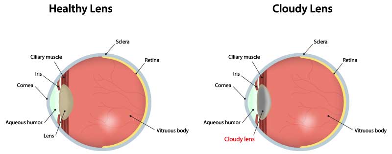
A cataract is a progressive clouding of the human lens inside the eye.
The lens is a small oval like structure and consists of a thin capsule (like saran wrap) enveloping a bag of protein. The protein is crystal clear and colorless at birth but discolors and clouds with age: first yellow, then brown, and finally cloudy. When the lens becomes cloudy and interferes with vision, it is called a cataract. The lens no longer transmits or focuses light clearly. A cataract is not a growth, a "skin", or a disease. It is usually not a sign of eye or systemic disease.
How does a cataract affect vision?
Initially the cataract changes the focusing power of the eye, and a change of glasses can satisfactorily improve vision.
Gradually, clarity drops so that visual tasks become increasingly difficult, despite the best possible glasses.
A person with cataract views the world as if through a dirty window or a windshield that needs defrosting. Glare becomes a serious problem. Color perception becomes muted.
Reading, driving, computer work, hobbies, and athletics become less easy and eventually impossible due to diminished vision.
What is Amblyopia? Page
Amblyopia is poor vision in an eye that did not develop normal sight in early childhood. It is often called "lazy eye". The condition is quite common, affecting 3 out of every 100 people.
Causes:
Amblyopia is caused by: Any abnormality that affects normal use of the eyes and vision development. Misaligned eyes, such as crossed eyes, often accompany amblyopia. Refractive errors (Needing eye glasses) are a frequent and treatable cause of amblyopia. Eye diseases, like cataracts, can lead to "lazy eye." Inherited conditions may also contribute to amblyopia.
Symptoms:
Blurred vision and/or crossed or wandering eyes is the major symptom of amblyopia.
Treatment:
Amblyopia MUST be diagnosed by age three in order for treatment to be the most effective. Primarily, the child must be forced to use the weak eye. A patch or cover is placed over the "good" eye for a prescribed number of hours daily. This is done for weeks, months or even years at a time.
Glasses are often prescribed to maximize the clarity of vision and vision development. Medication, such as drops, may be prescribed, or surgery may be required to remove cataracts or to straighten crossed or wandering eyes.
Treatments for the conditions that "cause" amblyopia do not cure it. Amblyopia must be treated in addition to the conditions that cause it.
What is a Detached Retina? Page
The retina is a thin, transparent tissue of light sensitive nerve fibers and cells, which covers the inside wall of the eye. When the retina becomes separated from the wall or tears, that part cannot work properly.
Symptoms:
Several symptoms are associated with detached or torn retinas and may include
- Blurred vision or blind spots and floaters
- Flashes of light
- Dark shadows in side vision or total vision loss
Causes:
Along with normal aging, detachments and tears may be caused by eye inflammations, complications from diabetes, tumors, injuries.
Treatment:
Surgery is the only treatment for detached or torn retinas and may be accomplished one of three ways.
Laser Photocoagulation:
Small burns which form scar tissue are placed around the edge of the tear to prevent fluid from passing through and collecting under the retina. No surgical incision is preformed.
Freezing (Cyropexy):
It is freezing the eye wall behind the retina to form scar tissue and seal the edges of the tear. Similar to the laser treatment but requires an anesthesia to numb the eye.
Surgical Repair:
Depending on the extent of the tear or detachment, the fluids might be drained from behind the retina and then the wall is pushed up against the retina. The amount of sight restored from a reattached retina varies depending on the length of time the retina was detached. Over 90% of all retinal detachments can be reattached successfully with modern surgical techniques.
How the Eye Works Page

How the Eye Works Anatomy of the Eye
Light enters the eye through the clear outer dome, the cornea, and goes through the pupil. The lens is just through the pupil: it transmits and focuses images on the retina, which is like film in a camera. The macula is the central receptor site of the retina and provides our sharp vision for tasks such as reading and driving. The retina sends the images via the optic nerve to the brain for visual processing.
Why We Don't See - Focusing Problems
Near-sighted or myopia The focusing elements of the eye are too strong and light comes to focusing front of the retina. The eye is "too long or too strong.
Far-sighted or hyperopia
The focus of the eye is too weak and the image of what we should see is not yet focused when it hits the retina. The eye is "too weak or too short."
Astigmatism
The eye is not perfectly round and the image of what we see is twisted or distorted.
Presbyopia
Occurs with all eyes: normal vision, myopia, hyperopia, and astigmatism. It is not a shape problem but is an internal aging problem. The eye muscles cannot change the shape of the lens to focus on near objects. Nearvision declines and reading glasses or bifocals become necessary.
Glaucoma Page
If you're one of the 1 million Americans who have been diagnosed with glaucoma... Consider yourself lucky! You've been given the opportunity to preserve your vision, because vision loss from glaucoma can be prevented if it is caught and treated in time. Many others are not so lucky. Almost 80,000 Americans are blind from glaucoma, and another million are at risk for vision loss because they don't know they have it.
In fact, glaucoma is one of the leading causes of preventable blindness in the U.S., and the single most common cause of blindness among African-Americans.
Glaucoma is often called the "sneak thief" of sight because the most common type causes no symptoms until vision is already damaged. That's why the best way to prevent vision loss from glaucoma is to know your risk factors and have medical eye examinations at appropriate intervals. (Your ophthalmologist can help you determine how often you should have your eyes examined.)
What is Glaucoma?
Glaucoma is a condition in which the optic nerve, which carries the images we see to the brain, is damaged. The optic nerve is like an electric cable containing about 1.2 million wires. Glaucoma can damage nerve fibers, causing blind spots to develop.

What causes glaucoma
Many people know that glaucoma has something to do with pressure inside the eye - the intraocular pressure (IOP).
Pressure builds up in the eye when the clear liquid called the aqueous humor, which normally flows in and out of the eye, is prevented from draining properly. This can happen in different ways, depending on the type of glaucoma. The resulting increase in pressure within the eye can damage the optic nerve.
Ophthalmologists used to think that high intraocular pressure was the main cause of optic nerve damage in glaucoma, however we now know that even people with "normal" IOP can experience vision loss from glaucoma -- so-called "normal tension glaucoma".
Some people with high intraocular pressure (also known as ocular hypertension) never develop the optic nerve damage of glaucoma. (These people need to be followed carefully by an ophthalmologist, because they are considered "glaucoma suspects.")
There may be other factors which affect the optic nerve, even when IOP is in so-called "normal" range. Elevated IOP is still considered a major risk factor for glaucoma, though, because studies have shown that the higher the IOP is, the more likely optic nerve damage is to occur.
If you think you're at risk for glaucoma, and haven't had a medical eye examination in the past two years, you can call us now for the Appointment that may save your sight.
Symptoms of Glaucoma
Most people who have glaucoma don't notice any symptoms until they begin to lose a significant amount of vision.
As optic nerve fibers are damaged by glaucoma, small blind spots may begin to develop, usually in the side -- or peripheral -- vision. The top photo at left shows how a scene would be viewed by a person with normal vision. The bottom image shows the same scene as viewed by a person with glaucoma. Many people don't notice the blind spots until significant optic nerve damage has already occurred. If the entire nerve is destroyed, blindness results.
One type of glaucoma, acute angle-closure glaucoma, does produce noticeable symptoms. In angle-closure glaucoma, there is a rapid buildup of pressure in the eye (intraocular pressure, known as IOP), which may cause any of the following:
- blurred vision
- severe eye pain
- headache
- halos (which may appear as rainbows) around lights
- nausea and vomiting
Angle-closure glaucoma is a rare, but serious, form of the disease. If you have any of these symptoms, call your ophthalmologist immediately. Unless treated quickly, blindness can result
Diabetes and Your Sight Page
Diabetes is a leading cause of vision loss in this country and around the world. This loss of vision is largely preventable with early screening and treatment. If you have diabetes, your body does not use and store sugar properly. High blood-sugar levels can damage blood vessels all over your body, including your eyes. Diabetes damages blood vessels in the retina, the layer which receives and transmits visual images to your brain. This damage is called diabetic retinopathy.
Types of Diabetic Retinopathy
Diabetic changes in the retina develop in stages, on a continuum. These stages are divided into early, nonproliferative, changes, referred to as background retinopathy, or BDR; and more advanced, proliferative, changes, or PDR.
In background retinopathy, or BDR, early damage to blood vessels leads to leaking of blood and fluid into the retina. If there is enough leakage the retina will swell, decreasing vision. As BDR progresses, vision may also be lost due to tissue damage from inadequate blood supply.
In more advanced or PDR, abnormal new vessels begin to grow in the retina. These vessels do not supply normal blood flow to retinal tissue. As the vessels grow they may cause severe bleeding, retinal detachment and glaucoma, all leading to significant loss of vision.
Diagnosis and Treatment
Regular exams by an ophthalmologist can detect early changes and help prevent vision loss. If significant disease is found, retinal photography and other tests may be necessary. Background retinopathy which threatens or affects vision with retinal swelling can be treated in the office with short laser treatments. This will greatly reduce the risk of visual loss. More advance disease is also treated with laser, often in multiple sessions. Bleeding or retinal detachment may require more involved surgery.
Vision Loss Is Largely Preventable
Early detection and treatment can reduce your risk of vision loss by a very large margin. With today's advanced diagnosis and treatment, only a small percentage of people who develop diabetic retinopathy have serious vision problems. Although strict sugar control reduces your risk of developing diabetic retinopathy, some patients with early or well controlled diabetes do develop retinopathy.
Early detection of diabetic retinopathy by an ophthalmologist is best protection against loss of vision.
When to Schedule an Exam
People with diabetes should see their ophthalmologist at least once a year. More frequent exams may be needed if retinopathy is detected.
Pregnant women with diabetes should schedule an appointment in the first trimester. Retinopathy can develop and progress quickly during pregnancy.
If you need new glasses, be sure your sugar is stable for at least several days before your appointment. Glasses that work well with an elevated sugar will not work well when sugar is stable.
Other Refractive Procedures
Clear Lens Extraction
Clear Lens Extraction or CLEAR for short is another vision correction option designed especially for people who are over 50 and are not good candidates for LASIK. Unlike LASIK and other refractive surgery techniques that correct vision by changing the shape of the cornea, CLEAR corrects your vision by changing the focusing power of the eye’s lens.
To do this, we replace your natural lens with a new intraocular lens (IOL). Both cataract surgery and Clear lens extraction have the option to use the Restore lens, which can restore distance, intermediate and near vision. Crystalens is an accommodating lens it acts like a natural lens by changing focus to allow distance, intermediate and near vision. These are the only refractive procedures that allow a full range of vision. While CLEAR is not right for everyone, it may be the option you are looking for. To find out if you are a candidate contact us about having an evaluation today.
- eye dr
- Eye Doctor
- eye dr florida
- eye fl
- LASIK Eye Doctors in Dora
- LASIK Eye Doctors in Clermont Florida
- Eye Specialists of Florida
- Eye care specialists
- lasik procedure
- Ophthalmologist
- eye care procedures in florida
- vision correction
- eye exams
- CLEAR
- Clear Lens Extraction
- Restore Lens
- Cataract Surgery
- Crystalens